
चित्र:Main symptoms of diabetes.svg

मूल चित्र (SVG फ़ाइल, साधारणतः 1,029 × 1,052 पिक्सेल, फ़ाइल का आकार: 863 KB)
|
|
यह फ़ाइल विकिमेडिया कॉमन्स से है। वहाँ पर इसका विवरण पृष्ठ निम्नोक्त है। कॉमन्स मुक्त लाइसेंसों के अंतर्गत उपलब्ध मीडिया फ़ाइलों का संग्रह है। आप भी इसमें मदद कर सकते हैं। |
सारांश
| विवरण |
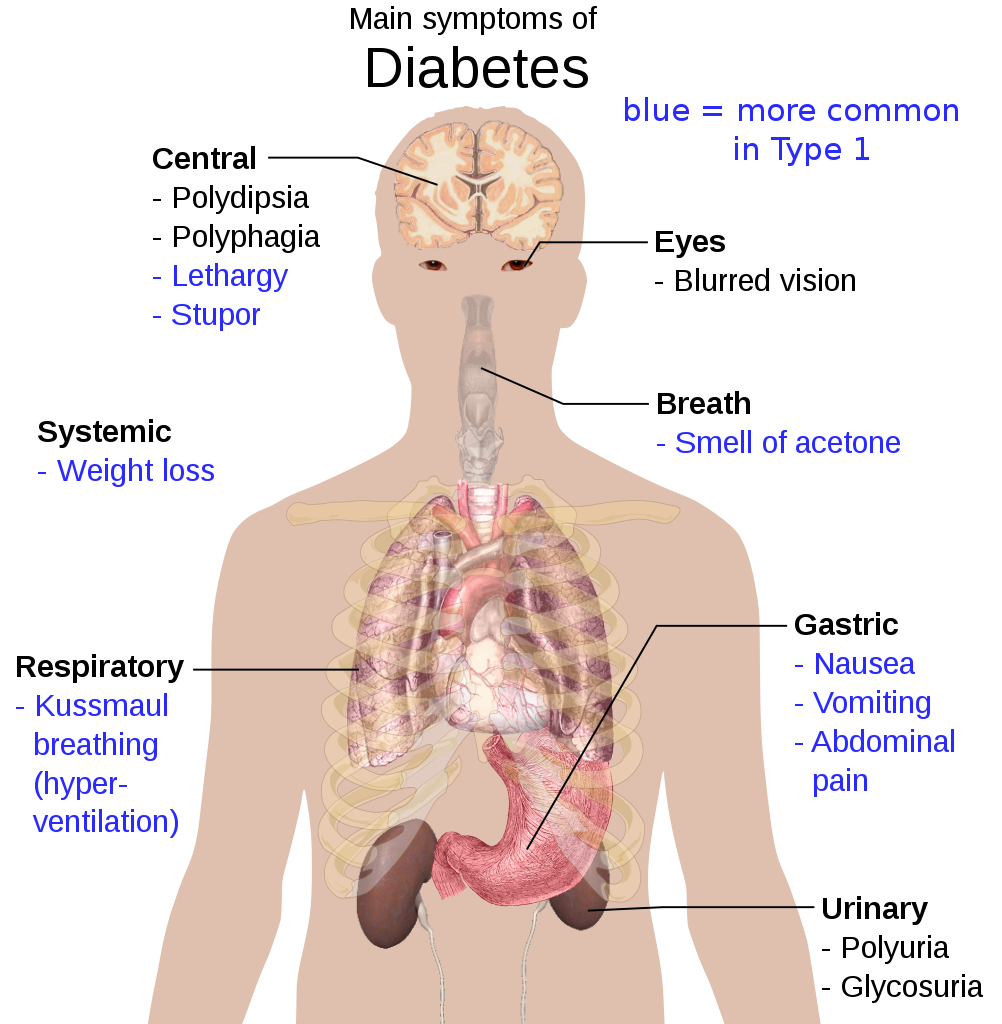
English: Overview of the most significant possible symptoms of diabetes. See Wikipedia:Diabetes#Signs_and_symptoms for references.
To discuss image, please see Template talk:Human body diagrams
Español: Principales síntomas de la Diabetes. Azul: más común en Tipo 1. Central: Polidipsia, Polifagia, Letargia, Estupor. Sistémico: Pérdida de peso. Respiratorio: Respiración de Kussmaul (hiperventilación). Ojos: Visión borrosa. Aliento: Fetor cetónico. Gástrico: Náuseas, Vómitos, Dolor abdominal. Urinario: Poliuria, Glicosuria. |
| दिनांक | |
| स्रोत | See above. All used images are in public domain. |
| लेखक | Mikael Häggström |
| दूसरे संस्करण |
[]
|


.png)


|
इस SVG फ़ाइल में एम्बेड किया हुआ टेक्स्ट है जिसे आप अपनी भाषा में किसी भी सक्षम SVG एडिटर, टेक्स्ट एडिटर, या फिर SVG Translate उपकरण की मदद से अनुवादित कर सकते हैं। अधिक जानकारी के लिए देखें: SVG फ़ाइलों को अनुवादित करने के बारे में। |
लाइसेंस
| मैं, इस कार्य का/की कॉपीराइट धारक, इस कार्य को सार्वजनिक डोमेन में प्रकाशित करता/करती हूँ। यह पूरे विश्व में लागू होता है। कुछ देशों में यह कानूनी तौर पर नहीं हो सकता है; ऐसा हो तो: मैं सभी को इस कार्य का इस्तेमाल किसी भी उद्देश्य से, बिना किसी बाधाओं के इन शर्तों के कानून द्वारा अनिवार्य किए तक करने की अनुमति देता/देती हूँ। |
Other versions
-
 hrvatski
 -
 español (spanish)
 -
 macedonian
Human body diagramsMain article at: Human body diagrams Template location:Template:Human body diagrams How to derive an imageDerive directly from raster image with organsThe raster (.png format) images below have most commonly used organs already included, and text and lines can be added in almost any graphics editor. This is the easiest method, but does not leave any room for customizing what organs are shown. Adding text and lines: Derive "from scratch"By this method, body diagrams can be derived by pasting organs into one of the "plain" body images shown below. This method requires a graphics editor that can handle transparent images, in order to avoid white squares around the organs when pasting onto the body image. Pictures of organs are found on the project's main page. These were originally adapted to fit the male shadow/silhouette.
Organs:
Derive by vector templateThe Vector templates below can be used to derive images with, for example, Inkscape. This is the method with the greatest potential. See Human body diagrams/Inkscape tutorial for a basic description in how to do this.
Examples of derived works
Licensing
|


.png)


चित्र का इतिहास
फ़ाइलका पुराना अवतरण देखने के लिये दिनांक/समय पर क्लिक करें।
| दिनांक/समय | थंबनेल | आकार | सदस्य | प्रतिक्रिया | |
|---|---|---|---|---|---|
| वर्तमान | 20:40, 11 मई 2020 | | 1,029 × 1,052 (863 KB) | Goszei | Removed grey box around blue-text note |
| 07:35, 20 जनवरी 2012 |  | 1,029 × 1,052 (863 KB) | Mikael Häggström | Updated | |
| 18:13, 27 फ़रवरी 2009 |  | 511 × 565 (346 KB) | Mikael Häggström | {{Information |Description={{en|1=g}} |Source=g |Author=g |Date=g |Permission= |other_versions= }} <!--{{ImageUpload|full}}--> |
चित्र का उपयोग
इस चित्र से कोई पन्ने नहीं जुड़ते
चित्र का वैश्विक उपयोग
इस चित्र का उपयोग इन दूसरे विकियों में किया जाता है:
- bcl.wikipedia.org पर उपयोग
- bn.wikipedia.org पर उपयोग
- bo.wikipedia.org पर उपयोग
- en.wikipedia.org पर उपयोग
- es.wikipedia.org पर उपयोग
- eu.wikipedia.org पर उपयोग
- fa.wikipedia.org पर उपयोग
- ga.wikipedia.org पर उपयोग
- he.wikipedia.org पर उपयोग
- hy.wikipedia.org पर उपयोग
- jam.wikipedia.org पर उपयोग
- ja.wikipedia.org पर उपयोग
- ko.wikipedia.org पर उपयोग
- mnw.wikipedia.org पर उपयोग
- ms.wikipedia.org पर उपयोग
- nia.wikipedia.org पर उपयोग
- sl.wikipedia.org पर उपयोग
- sq.wikipedia.org पर उपयोग
- sv.wikipedia.org पर उपयोग
- vi.wikipedia.org पर उपयोग